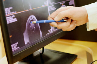
Investigations
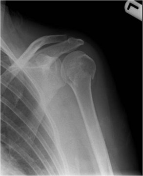
As part of your consultation, you may be required to have a plain X-Ray. This gives us good information about the bones and joints but does not give us specific information about soft tissues.
Below shows a plain X-Ray of the shoulder.
What types of scans are there?
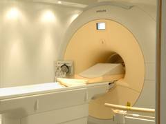
There are several types of more detailed scan, but the most commonly used is Magnetic Resonance Imaging – MRI. This uses a large magnet to create the images and gives detailed pictures showing the anatomy of both bones and soft tissues.
A MRI scanner is shown below.
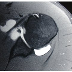
MRI shows abnormalities of ligaments, rotator cuff tendons and labrum of the shoulder very well. There are certain structures in the shoulder which can be seen better following an injection of contrast into the shoulder (arthrogram) your surgeon will guide which type is required. Patients who suffer from claustrophobia may have difficulty with having an MRI scan.
Below shows an MRI arthrogram of the shoulder.
Other less frequently used scans are CT (computerised tomography). It is best for showing the anatomy of bones, it is still frequently used to assess fractures, allowing description of the fracture fragments, 3 dimensional reconstruction is now available to recreate the anatomy of a fracture or post injury deformity. It is also useful for determining bone loss in the socket of the shoulder in patients with shoulder arthritis or shoulder instability
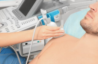
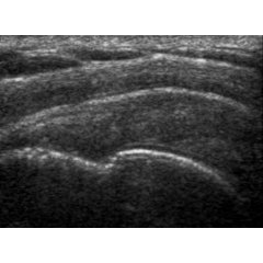
Ultrasound is also used in shoulder surgery, it is used to look at the rotator cuff tendons. Some injections into the shoulder can also be given with Ulrtasound guidance.
Below shows an ultrasound of the shoulder being done, looking for any abnormality in the rotator cuff.
Andrew Brooksbank

CONSULTING HOURS
CONTACT DETAILS
Treatment journey
APPOINTMENTS
Make a consultantion appointment wiith Mr Andrew Brooksbank at BMI Ross Hall Glasgow.
FAQ’s
Frequently asked questions about appointments, treatment, recovery and insurance/payments.
